AV Block ECG Patterns
Learn to recognise AV block ECG patterns by assessing the PR interval, dropped QRS complexes and the relationship between atrial and ventricular activity. Clinical impact depends on the degree of block, the resulting ventricular rate, and perfusion.
Quick Jump
P ↔ QRS relationshipOverview
PR + dropped beatsThe term “AV block” describes conduction abnormalities between the atria and ventricles. These can range from a simple delay (first-degree block) to intermittent failure of conduction (second-degree block) to complete dissociation (third-degree block). ECG interpretation focuses on the relationship between P waves and QRS complexes, and on the PR interval.
First-Degree AV Block
Delay onlyFirst-degree AV block is a conduction delay (not a “dropped beat”). Every atrial impulse conducts to the ventricles, but more slowly than normal.
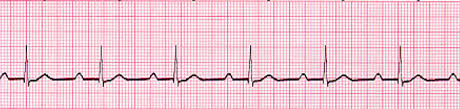
ECG features: PR interval is > 200 ms and constant across beats. P waves are followed by QRS complexes in a 1:1 pattern. QRS complexes are usually narrow unless there is additional conduction disease.
Second-Degree AV Block Type I (Mobitz I / Wenckebach)
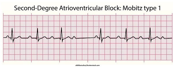
PR lengthensType I second-degree AV block is characterised by progressively increasing conduction delay until an impulse fails to conduct and a ventricular beat is dropped.
ECG features: PR interval progressively lengthens beat-to-beat until a non-conducted P wave occurs (a “dropped” QRS). After the dropped beat, the cycle repeats. The conduction abnormality is often within the AV node.
Second-Degree AV Block Type II (Mobitz II)
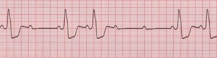
Dropped beatsType II second-degree AV block occurs when conduction fails intermittently without prior progressive PR prolongation. Some atrial impulses conduct normally while others do not conduct at all.
ECG features: PR interval is typically constant for conducted beats, but intermittent non-conducted P waves occur (dropped QRS complexes). This often appears in a pattern (e.g., 2:1, 3:1). The block is more likely within the His-Purkinje system and may be associated with a wider QRS if bundle branch disease is present.
Third-Degree AV Block (Complete Heart Block)
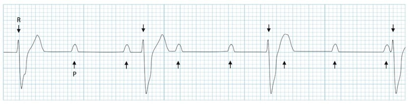
AV dissociationThird-degree AV block is complete failure of conduction from atria to ventricles. The atria and ventricles beat independently: atrial activity is driven by the SA node, while the ventricles are driven by an escape rhythm.
ECG features: AV dissociation with P waves and QRS complexes occurring regularly but with no consistent relationship between them. Ventricular rate depends on the escape focus: junctional escape rhythms are often faster than ventricular escape rhythms, and QRS width may vary accordingly.