ECG Arrhythmias: Sinus, Atrial and Junctional Rhythms
Learn ECG rhythm recognition for common sinus, atrial and junctional arrhythmias, including atrial fibrillation, atrial flutter, PACs and junctional rhythms.
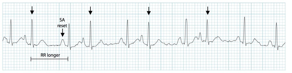
Sinus arrhythmia is an irregular rhythm caused by pacemaker cells in the SA node firing with variation.
The heart rate generally remains within normal limits, and the irregularity often corresponds with the respiratory cycle
(respiratory sinus arrhythmia).
During inspiration, increased venous return to the right heart is associated with a reflex reduction in vagal tone,
which can increase the sinus rate; ECG complexes may appear closer together.
During expiration, vagal tone may increase, slowing the sinus rate and producing a widening of the R–R intervals.
This is commonly benign and often produces no symptoms.
Sinus Bradycardia
Rate < 60
Sinus bradycardia is a sinus rhythm with a rate less than 60 beats per minute (in adults), typically regular.
It can be normal when metabolic demand is reduced (e.g., sleep) or in trained athletes.
Clinical significance depends on perfusion and symptoms. If associated with signs of poor perfusion, it is considered
symptomatic bradycardia.
Sinus Tachycardia
Rate > 100
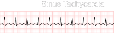
Sinus tachycardia is a sinus rhythm with a rate greater than 100 beats per minute. It is commonly driven by an underlying
physiological stressor (e.g., exertion, anxiety, pain, fever, dehydration, haemorrhage).
Sinus Pause and Sinus Arrest
Missing beat
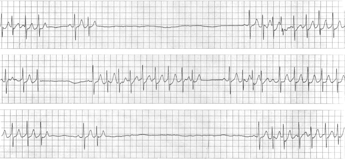
A failure of impulse generation from the SA node can result in a pause where an expected atrial depolarisation does not occur.
On ECG, this may appear as a “missing” P wave and associated QRS complex (a missing beat).
Terminology varies, but a brief interruption is often described as a sinus pause, while longer interruptions may be described as
sinus arrest. Recurrent or prolonged pauses can cause presyncope/syncope.
Sick Sinus Syndrome
SA node dysfunction
Sick sinus syndrome is a collection of SA node dysfunction patterns caused by abnormal impulse formation and/or impaired conduction
from the SA node to the atria.
It can present with sinus bradycardia, sinus pauses/arrest, and sinoatrial block, sometimes alternating with atrial tachyarrhythmias
(e.g., atrial fibrillation or flutter).
Atrial Arrhythmias
Supraventricular
Premature Atrial Contractions (PACs)
Early P
PACs originate from an ectopic atrial focus outside the SA node. The ectopic focus depolarises the atria prematurely, producing an
early P wave.
On ECG, the premature P wave often has a different morphology from the sinus P wave. The QRS complex is usually narrow and similar
to the underlying rhythm, and the PAC may be followed by a brief pause.
Atrial Tachycardia
150–250 bpm
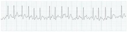
Atrial tachycardia is a supraventricular rhythm with impulse origin in the atria (not the SA node). Rates are commonly in the
range of approximately 150–250 beats per minute.
P waves may be abnormal in shape and may be difficult to see (sometimes merging with the preceding T wave). QRS complexes are
typically narrow unless there is aberrant conduction or pre-existing bundle branch block.
Atrial Flutter
Sawtooth
Atrial flutter is a supraventricular tachyarrhythmia commonly associated with organised atrial activity around a re-entrant circuit.
The atrial rate is often approximately 250–350 beats per minute.
On ECG, atrial activity may appear as “sawtooth” flutter waves (F-waves). The AV node typically conducts only some impulses
(e.g., 2:1, 3:1), so the ventricular rate is slower than the atrial rate.
Atrial Fibrillation
Irregularly irregular
Atrial fibrillation is chaotic, disorganised atrial electrical activity. Atrial rates can be very rapid (often described as
350–600+ impulses per minute), resulting in ineffective atrial contraction.
On ECG there are no discrete P waves. The ventricular response is typically irregularly irregular, because the AV
node variably conducts impulses to the ventricles. The baseline may show fibrillatory (f) waves.
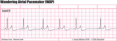
Wandering Atrial Pacemaker
Variable P
A wandering atrial pacemaker is an irregular rhythm where the pacemaker focus shifts between the SA node and other atrial sites.
This produces P waves with varying morphology and variable PR intervals from beat to beat.
Junctional Arrhythmias
AV junction
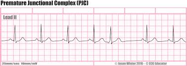
Premature Junctional Contraction (PJC)
Early beat
A premature junctional contraction originates from an irritable focus in the AV junction and occurs earlier than the next expected
sinus beat. Ventricular activation is typically normal (narrow QRS), but timing is premature.
If atria are depolarised retrograde, the P wave may be inverted (often in inferior leads), may occur before the QRS with a short PR,
may be hidden within the QRS, or may appear after the QRS.
Junctional Escape Rhythm
40–60 bpm
When higher pacemakers fail (e.g., SA node dysfunction), the AV junction can assume pacemaker function. A junctional escape rhythm
typically has a rate around 40–60 beats per minute and is usually regular.
P waves may be absent, inverted, or occur after the QRS due to retrograde atrial depolarisation. QRS complexes are typically narrow.
Accelerated Junctional Rhythm
60–100 bpm
Accelerated junctional rhythm occurs when the AV junctional rate is faster than its usual escape rate, commonly around 60–100 beats
per minute. P waves may be absent or inverted depending on retrograde conduction; rhythm is usually regular and QRS is typically narrow.
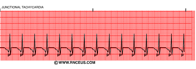
Junctional Tachycardia
100–200 bpm
Junctional tachycardia is a tachyarrhythmia arising from the AV junction, commonly with rates around 100–200 beats per minute.
P waves may be inverted and can occur before, during, or after the QRS complex. QRS complexes are usually narrow unless there is
aberrant conduction or pre-existing conduction disease.
Next: ventricular rhythms — or jump back to the cardiac index.