
Ventricular Arrhythmias on ECG
Recognise ventricular arrhythmias on ECG, including PVCs, ventricular escape rhythms, monomorphic and polymorphic VT, torsades de pointes and ventricular fibrillation. (Interpretation should always be combined with clinical context.)
Quick Jump
Find the rhythm fastPremature Ventricular Contractions (PVCs)
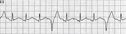
Ectopic beatA premature ventricular contraction (PVC) is an ectopic ventricular beat that occurs earlier than the next expected sinus beat. PVCs arise from a ventricular focus (below the AV node), so ventricular activation does not follow the normal His-Purkinje pathway.
PVCs can occur as isolated beats or in recurring patterns such as: bigeminy (every other beat is a PVC) or trigeminy (every third beat is a PVC). PVCs are often benign, but frequent PVCs or runs of ventricular beats can occur in structural heart disease and may precede sustained ventricular tachyarrhythmias.
ECG features: PVCs are typically wide with a bizarre QRS morphology, usually no preceding P wave for that ectopic beat, and they may be followed by a compensatory pause (leading to an irregular overall rhythm).
Idioventricular Rhythms and Ventricular Escape Rhythm
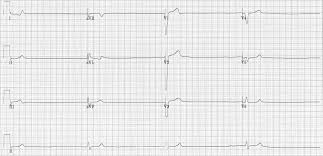
Last resortWhen higher pacemakers fail to generate or conduct impulses, a ventricular focus can initiate an escape rhythm. A ventricular escape rhythm is often considered a “rhythm of last resort” and typically occurs at a slow rate.
Typical rate: around 20–40 bpm (ventricular escape rhythm). A faster form, often called accelerated idioventricular rhythm (AIVR), is usually around 50–110 bpm.
ECG features: usually absent or dissociated P waves, wide QRS, and the ST-T may appear discordant (T wave opposite the main QRS direction), which can be a normal “secondary” repolarisation change in wide-complex rhythms.
Ventricular Tachycardia (VT)
Wide-complex tachyVentricular tachycardia is a ventricular rhythm with a rate typically ≥ 100 bpm and three or more consecutive ventricular beats. VT can be non-sustained (self-terminating) or sustained.
VT is potentially unstable because it can significantly reduce cardiac output and can deteriorate into ventricular fibrillation, particularly in the presence of structural heart disease or myocardial ischaemia.
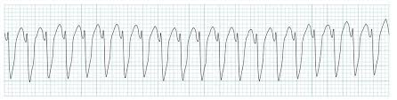
ECG features: usually a wide-complex tachycardia. The exact appearance depends on the VT subtype.
Monomorphic VT
Monomorphic VT has a consistent QRS morphology from beat to beat (uniform shape), suggesting a single dominant ventricular focus or stable re-entrant circuit.
Polymorphic VT
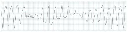
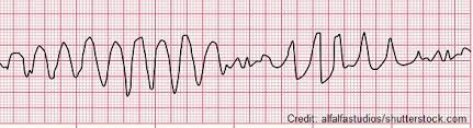
Polymorphic VT shows varying QRS morphology (shape changes beat to beat), suggesting changing ventricular activation. A classic form is Torsades de Pointes, which has a distinctive “twisting” QRS axis pattern and is often associated with prolonged repolarisation (prolonged QT/QTc) in the right context.
Ventricular Fibrillation (VF)
No outputVentricular fibrillation is chaotic ventricular electrical activity with no organised ventricular contraction. This results in no effective cardiac output. Without prompt intervention, it leads to death.
ECG features: a disorganised waveform with no identifiable P waves, QRS complexes, or T waves. The tracing may appear “coarse” or “fine,” depending on amplitude.
