
12-Lead ECG Lead Placement and Basics
A practical guide to 12-lead ECG lead placement, what each lead represents, how the leads are derived, and why correct setup matters before interpretation begins.
Quick Jump
Basics first → interpret secondWhat a 12-Lead ECG Represents
12 views • 10 electrodes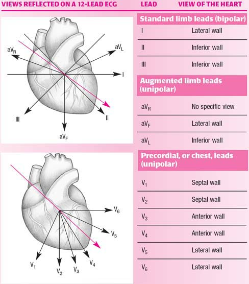
A standard 12-lead ECG provides 12 different views of the heart’s electrical activity using 10 electrodes placed on the body. Each “lead” represents a specific electrical viewpoint rather than a physical wire.
By combining information from multiple leads, clinicians can assess rhythm, conduction, chamber involvement, and patterns of ischaemia or injury with greater localisation than a single-lead ECG.
The 12 Leads
Grouped for sanity
The standard 12 leads are grouped as follows:
- Three bipolar limb leads: I, II, III
- Three augmented limb leads: aVR, aVL, aVF
- Six precordial (chest) leads: V1, V2, V3, V4, V5, V6
Although there are 12 leads, only 10 electrodes are required: four limb electrodes and six chest electrodes. The augmented limb leads are mathematically derived from the limb electrodes.
Frontal Plane (Limb Leads)
I, II, III, aVR, aVL, aVFThe limb leads (I, II, III, aVR, aVL, aVF) view the heart in the frontal (coronal) plane. These leads are particularly useful for assessing:
- Heart rate and rhythm
- Electrical axis
- Inferior and high-lateral territory changes
Horizontal Plane (Precordial Leads)
V1–V6The precordial leads (V1–V6) view the heart in the horizontal (transverse) plane.
- V1–V2: septal / right ventricular perspective
- V3–V4: anterior perspective
- V5–V6: lateral perspective
Why Correct Lead Placement Matters
Garbage in → garbage outIncorrect lead placement can significantly alter ECG appearance and may mimic or obscure pathology. Common consequences include:
- Apparent axis deviation
- False ST-segment elevation or depression
- Abnormal R-wave progression
- Misinterpretation of bundle branch block patterns
Positioning the Leads
Consistency mattersAccurate and consistent electrode placement is essential for reliable interpretation. The following video demonstrates standard precordial and limb lead placement.